Interpreting Mammograms: Calcifications

from the American College of Radiology Breast Imaging Boot Camp
Comments
-
The radiologist showed me the microcalcifications on the diagnostic mammogram. 1 spot was focused on. She told me that I had Stage 0 breast cancer and emphasized it was the earliest Stage to be caught. I am scheduled for a biopsy at a breast center. How accurate is the radiologist diagnosis, and can they see microinvasions? I'm very nervous.
0 -
Being sent for steriotactic biopsy, birad 4 b, 4 mm ductal carcinoma in situ. Radiologist said because of the way it's grouped and shaped he cannot even say it's any where near benign. Treatment would be surgery and radiation.. biopsy is to see if it's for certain cancer and if it's stayed in the duct or if it's invasive.
0 -
iris, I am confused by the combination of "BIRADS 4b" and the statement that the radiologist " cannot even say it's any where near benign", as the suspicion level associated with 4b is only "moderately low". The positive predictive value of BIRADS 4b is approximately 36%, meaning that about 36% of the findings with that classification will actually be bc. Can you post your imaging report here?
0 -
MTwoman,
Biopsy was done on the 21st results should be in on the 29th. Praying for benign
0 -
Hoping for b9 for you as well!
0 -
I am 63, was told by the mammographer I have calcifications. The radiologist never spoke to me, my doctor never spoke to me, the mammographer was the one telling me I had to do a magnification, then a test to see if I had enough tissue behind the nipple (where the calcifications are), then finally I need to come back for a biopsy. That was 3 weeks ago, My doctor never spoke with me, and when I called the office, I was told they don't do things that way. The doctor will only speak with me after the biopsy results are in. The radiologist making these decisions never showed her face to me. I felt like I was dealing with some Wizard of Oz, behind a curtain that no one could see.
Is this normal behavior from those that make these decisions? I feel that if I'm being told I need a biopsy, at least one doctor should explain to me exactly what they are seeing. Not that I don't have respect for mammographers, but they are not the ones making the decisions.
Frustrated.
0 -
TaraLeeOm-
That does sound incredibly frustrating! We're so sorry you're not getting the answers you need. Have you had the biopsy yet? Please keep us posted on what happens!
The Mods
0 -
I am getting the biopsy on the 26th. I almost feel like not getting it at this time, and finding anotherdoctor first.
0 -
Hi TaraLee,
I think you're doing the right thing by getting the biopsy. Once you do that, you can always find another doctor for a second opinion. A BiRads 4B is moderately high risk for malignancy. Better to know what you're dealing with. Best wishes to you and let us know how it goes!
0 -
TaraLeeOm - I hear you. Getting the runaround from the people you're entrusting your health with takes an emotional and physical toll - at a time when you are seeking calm and confidence. While I have generally liked the doctors and staff I've worked with at Emory's Winship Cancer Institute, I have had some issues with administrative snafus that I found unacceptable. I take advantage of every survey Emory sends me after a visit, and answer bluntly. I hope you get an opportunity to do so as well.
I'm glad you are getting your biopsy soon, too. Is it stereotactic?
And no, I do not believe it is normal behavior. You should go with a doctor you like and trust and have confidence in. You can be researching that now - you can always change! Meanwhile, you are not alone - we hear you!!!!!!!
0 -
hi i am 47 and had my first mamagram a couple of weeks ago and got called back due to still dense breast and went for the follow up and then
and then had a sounagram they said i calafictions and im having a biopy june the 4 im so anxious and i cant stay off the interent thanks for your help
0 -
I had calcifications for years and the radiologists never seem concerned. But when one area disappeared the radiologist was concerned. She said that invasive cancer "eats" calcifications. She herself told me that and did the biopsies. The area was so small that it turned out to be removed by the needle biopsy.
0 -
Hi there ,I've just had mamo and ultrasound, that showed calcification around old scar tissue from lumpectomy 7 yrs ago which had a 7mm density ,have no idea what's next onco is getting a second opinion becouse first radiologist couldn't say whether it was b9 or not so I'm not sure what will come next please help
0 -
It is very common for benign "dystrophic" calcifications to develop at the surgical site. The trick is to distinguish them from a recurrence as they are first beginning to form. Later on they develop a more classic benign appearance. Pretty sure they show up sooner than 7 years though (if in fact they are new this year). Choices are usually biopsy vs watch and wait. If they recommend a biopsy I would agree with that course of action. Did your initial diagnosis come from an investigation of calcifications?
0 -
Just saw this slide in an CME lecture, thought it might be of interest considering some of the questions I have seen from the group. It indicates the relative chance of calcifications indicating the presence of a malignancy by morphology (size and shape).
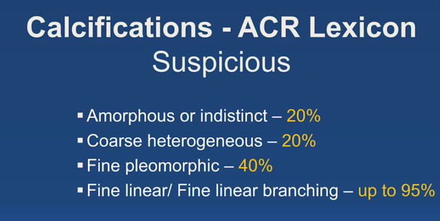 0
0 -
I'm confused on this, how can a radiologist even diagnose calcifications? I am scheduled for a biopsy on the 27th, 2 calcifications at about 11 and 12 o'clock and about 3CM and 1CM each.
0 -
I am not sure I understand your question.
0 -
This post.. Nov 12, 2017 11:51AM pamelakp wrote:
The radiologist showed me the microcalcifications on the diagnostic mammogram. 1 spot was focused on. She told me that I had Stage 0 breast cancer and emphasized it was the earliest Stage to be caught. I am scheduled for a biopsy at a breast center. How accurate is the radiologist diagnosis, and can they see microinvasions? I'm very nervous
that she is indicating that the radiologist said she had stage 0, how can a radiologist tell that on a mammogram without the biopsy? i'm just confused about that. I have had 2 mammograms but my biopsy is next week.
0 -
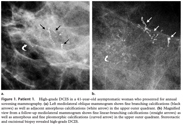
There are certain patterns of calcifications in the breast that are very recognizable and after a few years of training and a few years of experience one can make this call without hesitation. Unfortunately not all calcification patterns, or other findings in the breast, are as reliable as pathology in other parts of the body such as brain and bone tumors which can have very specific appearances lending themselves to predicting the histology of the tumor.
This pattern of branching microcalcifications, shown below, represents DCIS in the vast majority of cases. If there is no mass seen associated with it one assumes pure DCIS. By definition pure DCIS is stage 0 (still within the duct) but the DCIS with this pattern is usually high grade (grade 3). Also, no one will remove or treat any suspicious finding in the breast without a biopsy first.
 0
0 -
Thank you. I am still trying to figure out interpretation of mine I guess. I called back and the following day after my 2nd mammogram and they gave me the size which I said above and that it's about 11 and 12 o'clock, they didn't indicate any branching and said it was considered a loosely grouped coarse calcifications. I only have the 2, I also hadn't had a mammogram in 4 years so they didn't have anything recent to compare it to. My mom had stage 1 last year, IDC, I believe, had a lumpectomy and radiation so I'm concerned. I don't have my biopsy until the 27th so trying to stay positive. Any input?
0 -
Is your report posted anywhere on here? If not can you post it?
0 -
I didn't even get a report, only what they have told me on the phone.
0 -
should I call back and ask for something?
0 -
If they are new since your prior study and only on one side, and with a family history, a biopsy is reasonable. They will likely biopsy both groups but sometimes if the calcs are completely 100% identical in both groups and one is difficult to get to, they may only biopsy one group. Did your mother's present as calcifications or a mass? Picked up on a screener or felt by your mother or her doc?
Can you get your report through a patient portal?
0 -
No, not able to get my report that way, it would have been sent to my obgyn so I could call and ask for it. My mom said that hers was a tumor, was never identified as a mass or calcifications or anything like that, hers was found on a mammogram and same thing, sent for a biopsy.
0 -
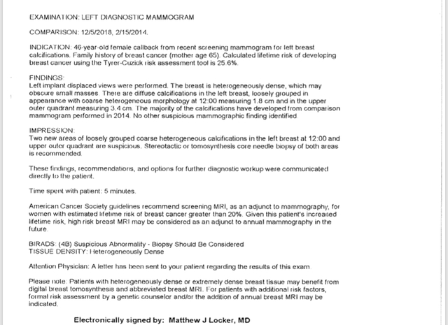
I got the report, it's a 4B
0 -
Can you copy or cut and paste it here?
0 -
 0
0 -
Any input? I'm of course freaking out now.
0