If you had DCIS with your IDC...
Comments
-
hi.
What started as DCIS stage 0 in my left breast has blossomed into Stage 1A, as it was found in the node they removed during mastectomy. So now we are looking at an entirely different ballgame. They are going back after the other nodes to see what's up. Hoping this doesn't suddenly indicate it is even more advanced than they thought. they say it is HER2, which is more aggressive? I'm having reconstruction. I haven't had the app't with the oncologists, radiologists yet to see what they are recommending. That comes next week. I am scared. Not of dying! Of the chemo and radiation. I know everyone's path is probably quite different...but am I looking at weeks of feeling sick and tired or months? I appreciate everyone's strength here....thank you all
0 -
Mimsy, I don't have experience with HER 2, I'm just jumping in to suggest you make a new post about this in the HER 2 board. or Stage 1, you should get helpful responses. I'm hoping for the best for you!
0 -
Mimsy - I answered you on the HDR2 threads. Probably a better place for you to be. But you've given better detail here. I didn't know you'd already has the mastectomy. Before any more surgery, you should meet with the MO - Medical Oncologist. Since chemo is likely, that should probably come next. Surgeon's cut - that's what they do. MO's become the driver of the bus once you have invasive disease.
Is not fun, but it's doable. You will be OK.
0 -
I had a biopsy in June 2020 that showed only DCIS. An MRI with contrast less than a month later did not change the doctor's opinion that it was still just DCIS, although quite a bit of it with all 4 quadrants of the breast involved. Had my double mastectomy in late August (I chose to do other side too - prophylactic). Even immediately after the surgery my doctor said he saw no evidence of anything other than DCIS. Full pathology was quite a shock. I had 26 "flecks" of Grade 3 invasive ductal. 4 lymph nodes were taken and while 3 were clean, one had what they called a "micro" amount of the grade 3 in it. So I am staged at 1A.
It took me a while to get through the tears and anger of why they didn't find the invasive sooner, or pushed for surgery sooner. But now I can accept that there is no point in the "what ifs". Having chemo, and trying to find some joy in each day.
0 -
Thanks all. I am new to all this and consider myself very lucky as it was only through my surgeons "magic fingers" and then my self-advocacy that my cancer was found as early as it was. The abnormal mammogram in Sept. 2020 that started everything resulted in a stereo-tactic biopsy (that was "fun") that indicated benign fibrocystic "stuff", but the pre-biopsy exam by my surgeon identified a palpable lump (5 o'clock) in the right breast that was missed by the mammo (my breast tissue is super dense - guess that's the definitive explanation of "boobs") and all other exams (including self), which turned out to be DCIS grade 2 with microinvasion. Then the MRI that was intended to be used to pinpoint surgery on the right identified IDC in the left breast (11 o'clock). The MRI was originally scheduled for 2 days before lumpectomy surgery but I asked if it could be scheduled earlier just incase the left breast was "hiding" something too. My wonderful care team was able to get it done on 10/19 and then hasten the recommend biopsy for 10/21 and schedule the bi-lateral lumpectomies with sentinel node biopsies for 10/23. Whew. Again, very lucky to have caught this early, margins and lymph nodes clear. Did genetic testing due to the atypical existence of cancer in both breasts. This too came back clear. Now awaiting results of tumor testing to see if Chemo would be beneficial. In the meantime am expecting radiation, but don't yet know the extent. Have also been told that Tamoxifen will be recommended but I am extraordinarily wary of taking this. I already have ridiculous hot flashes, mood swings and anxiety/depression (for which I have taken Lexapro for many years) and I can't imagine these symptoms worsening. Not sure I could handle that, and I am very nervous about weight gain and hair thinning. Why can't it create body thinning and hair thickening? My body is as thick as I want it and my hair is already too thin. Haha. Anyhow, thanks for all being out there and sharing. I am interested in finding alternatives for Tamoxifen and will look for other posts about this. Cheers.
0 -
Congratulations on being this side of "finding out". I agree that it is hard to understand why the pathology was so much more serious that the surgeon expected. I applaud you as well for choosing not only to treat the "known" side aggressively, but also to take prophylactic steps with the other side.dd That's a huge decision and one that obviously was wise. You know your own body better than anyone and you need to find the support and strength to be your own advocate and champion. Again, congratulations and best wishes for increased strength, balance and joy each day.
0 -
This is something I was wondering myself. I was just diagnosed triple positive idc with dcis. If I am reading the pathology report correctly, 1 of four cores was IDC and the other three were dcis. Im probably having a lumpectomy on the 15th. Wondering how much of my 2.5 cm tumour is idc and how much is dcis.
I'll try to remember to update here when I get the pathology report
0 -
Redcanoe, once you have your lumpectomy, they will go thru the resected tissue. They should be able to tell you the extent of the IDC. That seems to be how it works. I myself, had around 6 mm of DCIS, but they found in pathology, a 3 mm IDC tumor. They couldn't tell exactly how much DCIS there was in mine tho, which I found odd, and which I asked about. MO said it can be hard to tell exactly. They did seem to know it was 3.2 mm of IDC, so I guess that was not in question at least. Hope this helps.
0 -
To add to kksmom's info, staging is done based on the size of the IDC. Whether you also have 1mm of DCIS or 10cm of DCIS doesn't matter and doesn't affect the staging. So finding out the size of the IDC is what's important.
Generally IDC grows in place, becoming an ever larger mass as the cells multiply. DCIS cells are stuck in the narrow milk duct, so as DCIS cells multiply, the area of DCIS tends to spread out. This is why it may be easier to size the IDC and is also why some people have areas of DCIS that are very large (I had over 7cm, spread over two locations).
0 -
This is so interesting. I was just diagnosed with a 4mm IDC and a 6cm surrounding area that the surgeon suspects is DCIS. It seems we won't know for sure until surgery. I was thinking these same thoughts on my ride home from genetic counseling today. Why is the DCIS area around the IDC? Eventually would that whole DCIS area be IDC. I'm brand new here so my terminology if prob off. So effing much to learn!
0 -
Jen, sorry you've been diagnosed. Yes, there is a lot to learn but if you read this board and ask questions, you'll pick it up quickly.
"Eventually would that whole DCIS area be IDC."
No, not likely. Most DCIS usually remains DCIS. But what happens is that in one area (or sometimes more than one area), a few of the DCIS cells under go a biological change and develop the ability to break through the wall of the milk duct. The cells that break through and move into the open breast tissue are now invasive cancer cells, and as they multiply, the invasive cancer grows. But the rest of those original DCIS cells that remain in the milk duct are still DCIS, and probably will always remain DCIS. Eventually if the cancer isn't removed, would more cells evolve and break through the duct? Maybe, but never all of them if you have such as large area of DCIS.
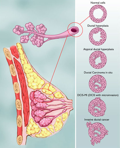
This image from BCO shows the process of cells development. You can see from the bottom two graphics that as the cells break through the duct wall, first as just a few cells (a microinvasion) and then as those cells outside the duct start to multiply, there still remain lots of DCIS cells in the duct.
 0
0 -
Thank you so much for your reply. I'm not digging too much on here until I know my hormone and genetic results so this helps me a lot. I'm very analytical so I'm proud that I've stayed here and haven't gone to Dr. Google. In case you haven't heard it lately, you are amazing with a big ole brain and have helped so many on here!
0 -
I remembered to update!
My final pathology was that there was 17mm of dcis within the 2.5 cm of idc. Which is interesting to me because when I had my original diagnostic ultrasound and mammogram, my tumour was 17 mm and very soft. Then after biopsy I felt it grow and become rock hard. I think maybe I caught it as it was starting to break out but unfortunately it took 7 weeks between my diagnostic mammogram and getting diagnosed and it was grade 3 triple positive cancer so it grew a lot. Cant help but wonder what the patho would have been if it was excised the day I found it.
0 -
Redcanoe,
It's not uncommon for an area that's had a needle biopsy to feel different, hard or swollen, after the biopsy.
DCIS is usually not palpable at all, so it is doubtful that what you were originally feeling was the DCIS. Certainly, a grade 3 triple positive tumor could be fast growing and could have doubled in size, or possibly even grown more, during the time between mammogram and surgery. But even the fasting growing tumor, with the possible exception of IBC, would not grow from a microinvasion to 2.5cm in 7 weeks.
How Fast Does Breast Cancer Start, Grow, and Spread? Breast Cancer Growth Rate and Doubling Time
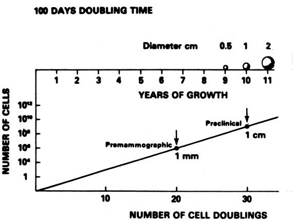
""Doubling time" is the amount of time it takes for a tumor to double in size. But it's hard to actually estimate, since factors like type of cancer and tumor size come into play. Still, several studies put the average range between 50 and 200 days." This article does mention one older study that suggested that the most aggressive tumors might double in as little as 25 days, but no other study has found a doubling time that is that short.This graphic isn't from the above study, but it provides an example of the change in tumor size based on doubling every 100 days. Obviously if a tumor is extremely aggressive and is doubling every 50 days, it will take much less time to reach 2cm in size, but we still are talking years not weeks.
 0
0 -
Hi Beesie,
Sorry for the silence..2020 was quite the year. I ended up getting a dbl mastectomy with reconstruction; implants. My body rejected the expander, on the radiated side, twice. So, I have decided to go flat, but even that won't be easy. I have an appointment with a new plastic surgeon next week, and I'm hoping what my general surgeon told me isn't the case. We shall see!
Have a great day!
0 -
I would love some more information about your dx, my pathology report seems to match yours pretty closely. Did you have lymph nodes involved? I'm trying to get educated about CA...I feel like every is jumbled in my head🤯
0 -
SLH, welcome to the discussion board.
You might want to start your own thread in the Just Diagnosed forum, with information about yourself (age is an important factor in treatment) and about your diagnosis. It's always confusing at first - we are thrown into a whole new world when we are diagnosed - and lots of us who've been through it can help you unjumble everything.
1 -
Hi Ladies
I am new in this forum. I was diagnosed Breast Cancer on August, 2020 , I had cancer with 2 stages on both of my breast. My left breast had stage0 DCIS 5cm (grade 3) and beside it there were stage 1B IDC with multi focal tumors ( grade 2) of 1.7cm , 0.8cm, 0.3cm, 0.2cm, 0.2cm. My right breast again I had stage0 DCIS 0.8cm ( grade 2) and stage1A IDC 0.9cm (grade 2). No lymph nodes involved.
I done all my treatments included of Bilateral Mastectomy, 4 chemos, 15 radiation and taking Tamoxifen. Right after my surgery my oncologist send my breast cancer sample to USA to get mamma print to see i am benefit from chemo. When the result come back with 19% risk of recurrence but I took chemos, radiation and Tamoxifen, it will bring down to 7% of risk of recurrence. I am very scare, I had 2 young kids 7 and 13. I am 46 this year. Anyone long term BC survivors whom had the same condition like my case , please contact me, give me some hope, support for this journey.
0 -
Hi My-Lan28, and welcome to Breastcancer.org!
We're so sorry for the reasons that bring you here, but we're really glad you've found us! You're sure to find our Community an amazing source of advice, information, encouragement, and support -- we're all here for you!
What a hand you've been dealt, for sure -- quite a lot of diagnoses to manage, but it sounds like you're on the tail end of things. Others will be by shortly to weigh in with their advice and experiences and give you the hope you're searching for. You may also want to start your own thread sharing your story in the Mixed Type Breast Cancer forum so that others can weigh in specifically on your situation.
We hope this helps and if you need any help navigating the forum, please let us know!
--The Mods
0 -
Mods, since most IDC develops from DCIS and ~80% of IDC diagnoses include some DCIS, to my understanding having both IDC and DCIS together in one diagnosis is not considered a "Mixed Type" breast cancer. It's in fact the most common presentation of breast cancer.
1