Interpreting Mammograms: Architectural Distortion
from the American College of Radiology Breast Imaging Boot Camp
(ligaments referred to are Cooper's Ligaments part of the support structure of normal breast tissue)
Comments
-
thanks DJ. Makes me feel good that I'm going for further evaluation
0 -
Really helpful post, as ever. Thanks DJMammo!
0 -
Thank you very much Dr. djmammo for the informative video...for some reason I never thought about looking up breast cancer information on YouTube and it is chock-full of helpful videos.
0 -
Thank you very much for posting this educational video.
0 -
TexasBelle06, we understand how scary this can feel. We tend to jump to the worst-case scenario.
Call-backs aren't unusual. It sounds like they are being thorough.
Take deep breaths, and distract yourself. And do let us know how things go.

The Mods
0 -
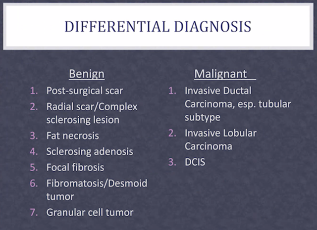
Here is a list of causes of architectural distortion both benign and malignant from a recent lecture I heard. They are listed in order of most common to least common causes.
 0
0 -
thank you for this! I do have a question. I am scheduled for biopsy next week for something very similar. Mine is at 1:00 posterior. I did have a breast reduction 15 years ago. Is it still possible for the asymmetrical AD showing up to be scar tissue?
Thanks!
0 -
It depends on whether or not its a new finding. If your current study was compared to a prior post-op study, and this finding is new, then it needs to be evaluated.
Also, in my experience the type of scarring present from reduction does usually not present as focal AD like a lumpectomy or other similar surgery.
0 -
Thank you! Unfortunately they had nothing to compare it to. I just had my first mammogram a few weeks ago and that’s when it was discovered.
The radiologist didn’t seem too convinced it was scar tissue, but I think he was trying to alleviate my concern
0 -
Jenney75
If they did an US, can you post it here
0 -
djmammo -
Thank you for being a valuable resource! I am 53 and due to very dense fibrocystic breasts have been having yearly mammos since I was 25 with no issues. I had an architectural distortion a few weeks ago. CNB showed stromal fibrosis but they want to excise entire area anyway - "just to be sure" rather than re-evaluating in 6 months - and refuse to discuss alternatives. I am an atty and that makes as much sense to me as telling a client who has not been charged with a crime that they should do a couple of months in county......just in case. Obviously I have a second opinion scheduled. My question in the meantime is whether the fact that they have had continued difficulty finding and "holding" the image of the distortion on ultrasound is meaningful. Even during the biopsy they had difficulty. All of the techs and radiologists were evasive when I questioned why it was so hard to consistently locate the suspicious area and how they could possibly determine how much to excise.
Again, many thanks for taking time out of your schedule to help women interpret their reports. I will return the favor by logging onto my Avvo account and answering some requests for legal help.
0 -
There are 3 causes of persistent architectural distortion on a mammogram. Scar from prior surgery, radial scar, and IDC. In a dense breast it can be a tough call to make. A cancer will usually show up on US, radial scar will but less reliably. Surg scar is accompanied by a hx of surgery. The cancers and the radial scars are removed.
If you can post the reports it might be easier to draw a conclusion.
Focal stromal fibrosis will often look like a cancer on US but is benign. When the report comes back from path, the radiologist will dictate an addendum to their report of the biopsy indicating the diagnosis and whether or not it is concordant with the imaging. If it doesn't match the initial impression, that is if it looks like cancer on imaging and path is benign, the findings are discordant and an excision is recommended. That is pretty standard everywhere. Sounds like thats what happened here.
When we perform an image guided core biopsy using 14, 16, or 18 g needles we make sure that we have 5 really good cores to send to path. This seems to be a fairly universal number set by the path departments everywhere I have practiced. We dont attempt to remove all of it.
0 -
Trudym if you don't get a comfortable answer please push for an MRI. My dense little breasts hid my cancer from standard imaging of mammography and ultrasound as well. While I eventually had an ultrasound guided biopsy, it was the targeted imaging of the MRI that allowed the doctor to determine the actual area to address. The mass just didn't show easily on ultrasound as it was close to the chest wall. I remember it took them over 20 minutes to agree where to begin the biopsy procedure. Wishing you the best.
0 -
Thank you so much for your response. The report is below. I have had no surgeries - just ridiculously fibrous dense breasts that I have had yearly mammos of for the past 25 years on advice of my GYN with no problems. I had inquired about either a second site needle biopsy or repeat mammo/ultrasound in 6 months to evaluate any change but both were immediately shut down. They are unable to tell me how much tissue would need to be removed until the surgeon was "in" as they could not get a consistently good picture of the distortion. It took forever for them to even locate it but they said it could be up to a bit bigger than a golf ball and, as it is at the top of my breast, I would likely need reconstruction. Everyone I spoke to seem almost offended that I was not immediately agreeing to surgery and provide only cursory responses to what I consider very basic questions that anyone would ask. With this kind of vagueness I am just not at all comfortable. My second opinion is scheduled for Monday but I am extremely grateful for your time and attention while I am waiting.
Addendum
Signed by Allen Jeffrey Levy, MD on 4/26/2019 1:51 PMADDENDUM:52-year-old female with history of left breast ultrasound-guided biopsy 04/24/2019. Pathology results have been finalized.
Final diagnosis:
Benign breast tissue with fibrocystic changes and cluster of small calcifications.
Biopsy results are discordant.
Recommendation:
Recommend breast surgical consultation for excisional biopsy of the suspicious region of architectural distortion.
Results and recommendation were discussed with patient. The duration of the consultation was 45 minutes of which more than 50% of the time involved counseling and coordinating care.
Patient is scheduled for breast surgical consultation with Dr. Theime on 05/16/2019 .
Study Result
Narrative
EXAM:
ULTRASOUND-GUIDED BREAST BIOPSY, LEFT
HISTORY:
The patient presents for ultrasound-guided biopsy a poorly defined hypoechoic mass in the left breast at 12 o'clock, corresponding with an area of mammographic architectural distortion.
COMPARISON:
US BREAST LIMITED LEFT dated 4/18/2019; MAMMOGRAM DIAGNOSTIC TOMO LEFT dated 4/18/2019; MAMMOGRAM SCREENING TOMO BILATERAL dated 4/15/2019.
FINDINGS:
The nature of the procedure, including the risks, benefits and potential alternatives, was reviewed with the patient, who gave her written consent to proceed. Images of the breast were obtained to localize the lesion in question. The overlying skin was then prepped and draped in the usual sterile fashion, and local anesthesia was achieved using a combination of 1 cc of 1% lidocaine solution and 10 cc of 1% lidocaine with epinephrine. A 12 gauge Suros ATEC biopsy device was inserted into the breast via a superolateral approach under direct ultrasound guidance. Five core specimens were obtained with vacuum assistance and sent for histologic analysis. A HydroMARK biopsy marker was deployed at the operative site. All needles were removed, and hemostasis was observed. The patient left the ultrasound suite with post procedure instructions in good condition.
A postprocedure mammogram demonstrates the biopsy marker at the expected operative site. There is no significant hematoma. There has been no other change.
IMPRESSION:
Uncomplicated ultrasound-guided biopsy of a hypoechoic mass at 12 o'clock in the left breast. Histologic analysis pending.0 -
Hi Rah -
Thank you so much for writing. I hope that you are feeling well! I have been thinking about an MRI and will definitely discuss one with the doctor I am seeing for a second opinion. I had an MRI about 10 years ago when I moved to PA and changed GYNs because she was appalled at the density of my breasts and wanted to make sure she had a good baseline on them. I can't say that laying on a table with the girls in holes was my favorite way to spend 20 minutes but we have to do what we have to do, right?
I suppose that is why this is throwing me for a loop. I have never had issues before despite yearly testing and have made sure I was cared for by professionals who treated me like an adult. Now I am being told that I need likely disfiguring surgery when there is no evidence of any actual problem by doctors who got defensive when I asked questions. Which is why I am no longer using the radiology/pathology/surgery practice my insurance company automatically shuttled me into and am using another practice that is still in network but apparently is not their "go to" people.
Thank you again for writing and wishing you health and happiness!!!
0 -
If the original report mentioned those calcifications as being in the area of arch distortion, they hit the target. This is further indicated by the followup mammo showing the marker in the same place as the original finding. A second image guided biopsy would likely show the same.
As it would be a shame to have an excisional biopsy of a wide area for what is potentially a benign condition, I agree that an MRI would give you the answers you need regarding what this is, and how to proceed from here.
If you are in PA, you could have your second opinion as well as your MRI at St Luke's Regional Breast Center in Bethlehem. It is a highly regarded diagnostic breast center. Where ever you go for the second opinion bring all your imaging on CD's and their reports along with the path report. The path slides could be sent out for second opinions as well. I know MD Anderson and Vanderbilt are sources for re-interpretation of the path slides. Keep us in the loop.
0 -
Thank you very much for taking the time to reply. I feel quite comfortable now that I am moving in the right direction by resisting pressure to have immediate surgery and seeking additional information before making such an important decision. I will definitely be asking for an MRI and ensuring that the physician I am seeing Monday has all of the information prior to my arrival. If for some reason this practice does not work out, Bethlehem is only a 3 hour drive from me.
Thank you again for donating your time. I can only imagine how many women your posts help every day.
0 -
Glad to help.
One more quick thing, if you do wind up having the surgery, find a surgeon whose practice is limited to breast procedures (rather than a general surgeon) and has training in oncoplastic surgery. They are the experts in a good cosmetic results in these cases.
0 -
Thank you - I definitely will. I am not ashamed to admit that cosmetic concerns are a major factor. There are not many benefits to infertility but the one major "consolation prize" is keeping your youthful figure and that includes maintaining breast shape and tone. If it is shown that there is a true likelihood of cancer then the scales tip on the risk/reward analysis but more information is needed.
BTW - Thanks for the inspiration to put in some extra time on Avvo. It is a good feeling and certainly got my mind off of all this!
0 -
Architectural distortion has got to be one of the most ambiguous and frustrating things to hear from a radiologist. I have been getting regular screening mammograms and 3D ultrasound screenings, due to the density of my breasts, religiously for the past 5 years. Last week I received a call to come back for a diagnostic mammogram and ultrasound. The ultrasound tech and the radiologist had discussion over whether they agreed on what they were seeing on the screen. There was no mass that could be seen. The radiologist asked me multiple times if I had surgery on the breast. The answer is no, but I had a very traumatic shoulder injury that resulted in surgery on the same side. The radiologist recommended an ultrasound guided core biopsy due to architectural distortion. I left the office extremely upset and very frustrated. My OBGYN was able to get me in for the biopsy on the 28th because she knows I'll drive myself crazy waiting 6 weeks for a biopsy appointment at the end of June which was the first date I was given. Both my mom and grandmother have had breast cancer. Luckily, both were diagnosed early and recovered. I am hoping if the biopsy shows cancer, that at least it was caught early.
0 -
I just had a lumpectomy last July for IDC upper rt quadrant of left breast. In 2000 I had a lumpectomy for dcis in the upper left quadrant with 6 weeks radiation. Call back mamo & US mamo report says architectural distortion. US report says lobulated mass with posterior shadowing at the 5 to 6 o'clock position.....
The Impression reads as follows: Suspicious mass subareolar left breast at the 5 to 6 o'clock position 1 cm from the nipple measuring up to 1.2 cm. This corresponds to an area of architectural distortion on the mammogram. This is separate from the post biopsy changes in the left breast. An Ultrasound-guided biopsy is recommended.
So what does this all mean?
lobular mass with posterior shadowing at the 5 to 6 left bwith reast, subtle
0 -
Yes biopsy is scheduled for May 31st. What does lobulated mass with posterior shadowing mean? Last year the US said with vascularity...but this one does not mention vascularity. So does that mean this mass does not have blood flow?
0 -
Lobulated is a description of the margin/border of the mass. More concerning than smooth margin, less concerning than irregular margin.
Posterior shadowing is an US specific finding indicating that the mass blocks sound rather than letting it go through the mass. It is a worrisome finding.
If a finding is not mentioned you cannot assume it was not present. They would have had to have said "no internal vascularity".
0 -
Dear DJmammo,
Thank you so very much for your time in answering my questions....it is fascinating and yet..concerning but I am scheduled for a biopsy and then when the results are in I will know for sure what it is and proceed from there. Thank you again for all you do on this site ~ to me, you are an earth angel!
0 -
Are there any lobules or ducts at the 5 to 6 oclock area?
0 -
The short answer is yes. Click this link:
0 -
Thank you again, DJmammo
 0
0 -
I have a lump that I was told was a fat lobule in 2014 and rechecked again in 2015. Now just was told I have a architectural distortion in that area and in one gland! Of course I am beyond scared, but wondering if that was misdiagnosed in previous years. Now the agony of waiting for the actual diagnosis. I feel with the gland being involved on mammogram it is pretty clear where this is going
0 -
Fat lobules and cancers causing architectural distortion look quite different. Besides if the fat lobule were a cancer it would have grown between 2014 and 2015. In the four years between then and now it would certainly be very large and palpable.
Not sure where you are in the process. Have you been recalled from a screening and waiting for additional mammo imaging and US or something else?
Not sure what is meant by "in one gland". Can you post the report(s)?
0