Lymph Nodes on Ultrasound
Comments
-
Quick update - I got more information during my ultrasound. The radiologist explained that the node in question was larger in comparison to my mammogram 1 year ago. She noted in ultrasound that it is enlarged and showed me the area. I would put it in the "a" diagram range - the white is still defined but the dark portion is larger than normal. I do not have reference for how enlarged it is but it is not palpable. They also did an ultrasound on my breast which still appears clear. Next up is punch biopsy on Thursday. Ugh, those hurt.
I have looked at my test results over the last 2 years and there may be some clues there which would not have stood out on there own but I will wait until the results of the biopsy due 3 days after the procedure and let you know. Also, the radiologist is the same who told me I most likely had breast cancer during round one so what the heck, bring on the flashbacks!
Thanks for being here, all. It really matters.
0 -
Verbal description (from my oncologist's nurse) of my path report for biopsy of my axillary node:
Tissue:
No evidence of malignancy
follicular lymphoid hyperplasia
Flow sytometry:
No evidence of lymphoma
No evidence of monoclonality
So, I'm good for now. I hope this information helps someone else! And whew, the stress valve really releases when you hear results and I, as now, had no idea how much pressure it was causing. Hang tough, all. Thank you for this thread, it was invaluable to have this space I trust to read other's expertise and experiences.
1 -
The chart posted above is very helpful - thank you djmammo. It is an appreciated resource when learning about what may possibly be growing. My 'mass' was first discovered in August 2018, had call back for dx mammogram and ultrasound. Went back in for 6 month check up. Have had a few look at images. It has been very confusing in that my mass has been classified as: possible node, complicated cyst, complex cyst, lymph node, not a cyst - solid - not sure, and possible intramammary lymph node. All agree that it is hypoechoic, has since become palpable (was not when first discovered), and seems to have established a 'feeding tail' - not previously seen. No one seems to be able to verify what it actually is - other than 'it is most likely benign' and wait a year for a recheck. There is something about no one being able to state what it most likely is with consistency and waiting a year that just makes me nervous. Some of my images look similar to the chart, but I am definitely no expert. Most that I talk with are shocked that no one thinks a biopsy is warranted.
I am curious if anyone else has had a lymph node issue. Did it seem to come out of 'no where'? Did anyone experience this 'vague' type of identifying what the growth is? Was it a lymph node or did it end up being something else? I know we have lymph nodes that swell and can get irritated/inflamed, but this intramammary term is new to me and I want to avoid heading to Google for information.
Any thoughts and/or insights would be greatly appreciated.
** Edit **
I am not sure if I am posting in the wrong areas, using language that does not gather feedback, or what. I just do not seem to get much if any feedback when I post. I did want to update and say that I am having a biopsy next week with a breast specialist. I'm sure all will be fine. It will be good to hopefully have an answer either way.
0 -
I’ve had a lymph node under my armpit on the good side for four years. It aches, I’ve had ultrasounds and another breast MRI and they say it’s normal but it feels like a marble to me. I just want it out. I’m so tired of worrying about it. They won’t do a biopsy. Is there anyone I can see just to have it removed?
0 -
Thank you Djmammo, I hadn't thought about that. Do you think that insurance may not pay for a biopsy if I push for it?
0 -
I’m Sorry DJmammo, it only allows me to send a certain number of private messages a day. To answer your private message: I went to the er bc I had pain and swelling. Pain and swelling is gone now. Yes you can feel the mass, it’s directly under my nipple. 2 days after my mammogram, I raised my arms and noticed an indention (dimple) on the same breast. But not even close to where my mass is. It’s towards the inside of my breast down close to the bottom. Also I had 2 separate occasions towards the end of last year where that same nipple discharged blood (without squeezing) no mass felt then. If my doctor thought it was a papilloma Why would she suspected it to be malignant? And why would there be increased vascularity? And give me a bi-RADS score of 4/5 from my ultrasound? Mammogram Bi-RADS 0 if i read the report correctly.
0 -
The ins company goes by the radiology report's recommendation. If the radiologist gives it a benign reading someone would have to call and advocate for you if you wanted a biopsy. Perhaps your PCP or ObGyn can order it on the basis of clinical findings and get it paid for that way.
0 -
Thanks Djmammo, I will see what they say next time I’m in for an appointment
0 -
Hello,
I'm new here. I don't know much yet. I went for a routine mammogram and was called back two days later to do an ultrasound. I went for my ultrasound and the report says that they are seeing a intrammamary nodule hypoechoic,with irregular margins and probably a fatty hilum. I will be contacted from the hospital for further assessment and maybe a biopsy but they are not sure yet. I don't understand. How can I have a malignant nodule if they cannot see any tumor. Worried sick. My doctor says I have nothing to worry about. Anybody else have thiskinda of results? Please reply.
0 -
Hi djmammo!
My lymph node US result has reniform shape with preserved fatty hilum but eccentric cortical thickness meausring 0.6cm , and second one has reniform shape with preserved fatty hilum uniformly thickness cortex measuring 0.6cm . what does this mean? Im so scared to death , fear of unknown
 . God bless whoever can decipher thi0
 -
Se answer to your PM.
0 -
if a lymph node has doubled in size to 3.5 cm over the past year and a half, but no other issues on the mammogram, would that be indicative of cancer? I have a biopsy on Monday, and I am on the verge of a panic attack with the worry.
0 -
The overall size can be misleading. The important measurement is that of the cortex of the node and the appearance of the central fat in the center of the node. These are best demonstrated on US.
You can have a 4cm node with a 2mm cortex and large amount of fat centrally and be benign. Alternatively you can have a 1cm node with a cortex so thick it compresses the central fat to a mere sliver and be malignant.
Important to note is that nodes can enlarge for many reasons that are completely unrelated to the breast and breast cancer. Also it would be unusual to have a node measuring 3.5cm due to the spread of a breast cancer without the primary tumor having already made its presence known.
0 -
thank you for the reply. I'm concerned that it has grown over the past year. Everything I'm reading says this is indicative of some sort of cancer.
0 -
My response reflects everything that I have read.
Post the report with the exact measurements taken of the node including the thickness of the cortex and the appearence of the fatty center, how many enlarged nodes were seen on that side and how many were seen on the other side. Are there any in your neck or groin? Have you had recent abnormal blood work?
Let us know what the biopsy shows.
Click here for more info: https://www.verywellhealth.com/axillary-lymph-nodes-2252131
0 -
the report I got is that they are calling it probably benign. It previously measured 2.6x1x1.1. Now it is 3.6x1.1x1.9. They state that it likely represents a lymph node. A surgical consult is recommended (I'm going Monday). I wasnt given any other details. They are closed today and nothing is showing in my online chart. I guess I just have to wait it out and pray for the best.
0 -
Hi everybody. Is it possible to have cancer in the lymph nodes even if they feel normal on palpation and appear normal on ultrasound? Mine appeared normal on u/s and were reportedly normal on palpation but following neoadjuvant chemo I had a positive node and 4 others that felt abnormal but were clear. I’m wondering if all5 were affected and 4 tested clear due to the chemo. Any insight on this? thanks
0 -
djmammo has provided a wonderful batch of information in the header. I'm bumping so new comers will be able to read his explanation when they are worried.
Yes, I had cancer in a lymph node that felt normal & did not show up anywhere - until it did.
0 -
They are not going to do a biopsy. She says it has clearly defined borders and looks normal. I'm to go back in 3 months to get it checked, and if it has grown, I will get a biopsy then.
0 -
djmammo,
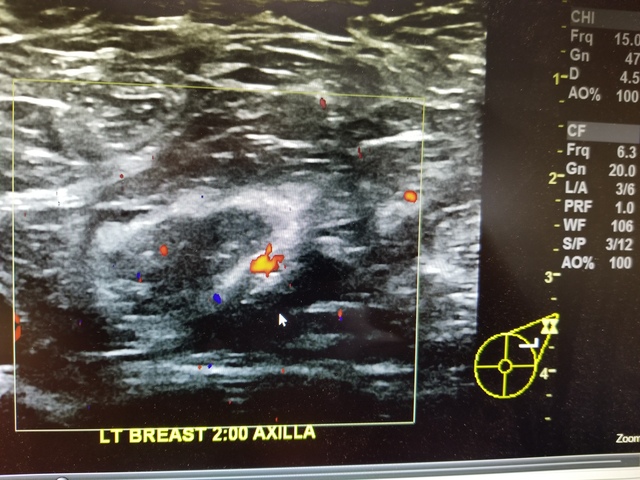
I'm getting the runaround as far as the thickness of the cortex and appearance of the fatty center. The breast specialist just tells me to look at the radiologist's report (and that info is not in there), and they tell me there is no way for me to talk to the radiologist directly. Are you able to tell anything from this ultrasound image?
0 -
Almost a year ago I began experiencing pain in my left breast. There was also a strange indented line forming when I lifted it with a small lump. I went in for a mammogram and received a letter a few weeks later saying that they wanted to do more imaging. I went back for another mammogram and was told it was just a lymph node. After the pain continued, I was eventually seen for an ultrasound and was told again it was just a lymph node.
Fast forward to today, and after experiencing continued pain coupled with a significant amount of weight loss, I was referred for another mammogram and ultrasound. This time around, they told me they didn't see anything. Not even the aforementioned lymph node.
I got the impression they felt I should be relieved, but in fact I'm feeling quite the opposite. A lymph node doesn't just disappear, does it? Shouldn't it have been relatively easy to find based on previous imaging? Shouldn't they have made sure to find it so they could compare it against previous imaging? I'm feeling very much like they didn't do what they were supposed to and I'm not really sure what to do about that.
0 -
The problem I had with a lymph node ultrasound, during my breast exam, was that nothing was shown. I had the US and MRI and everything looked to be clear.
A month later, 4 nodes were positive, one being 8mm. Terrifying that imaging cannot always detect these things as it may have changed my treatment plan.I likely would have had chemo before surgery to better understand how my lesion would have responded to chemo ...now I just don't know.
1 -
Hi DJMammo - sure! It is below. Post-surgery, my lesion was 5.5cm and a second was 1.7cm. 4/4 nodes were positive, but again, the lymph nodes were a big surprise.
Your insight is appreciated!
FINDINGS:
The breasts have extremely fibroglandular tissue. The background
parenchymal enhancement is mild.
LEFT BREAST: There is a vitamin E marker overlying the upper outer
posterior left breast. There is focal susceptibility in the upper outer
posterior left breast from a biopsy clip. There is an 8.6 x 5.3 x 4.8 cm
area of regional non-mass enhancement in the upper outer left breast,
which extends posteriorly to the chest wall and corresponds to the site of
biopsy-proven malignancy. The anterior extent of non-mass enhancement is
located on series 9/image 87 and series 20/image 55. There are mixed
kinetics with areas of rapid initial and washout delayed phase
enhancement. There is an indeterminate 1.3 x 0.6 x 1.4 cm oval
circumscribed enhancing mass in the central posterior left breast 9.3 cm
posterior to the nipple with rapid initial and persistent delayed phase
enhancement (series 9/image 111, series 20/image 54). There is an
indeterminate 0.6 x 0.4 x 0.5 cm area of clumped non-mass enhancement in
the central posterior left breast 7.4 cm posterior to the nipple with
rapid initial and persistent delayed phase enhancement, which is located
approximately the 1.0 cm anterior to the 1.3 cm mass (series 9/image 114,
series 20/image 57). Additional scattered foci of enhancement in the left
breast are favored to represent background parenchymal enhancement.
The left axilla is within normal limits.0 -
I wasn't given the reports, the tech just came back into the room after consulting the radiologist and said they didn't see anything and I could go. The breast specialist I'd seen previously later gave me a call and said they didn't see anything, not even the lymph node. I asked her if that meant it was smaller or just not visible because it confused me, and she said it was just not visible and that they don't always show up. Are the reports something I can request? I didn't realize. I have Kaiser and have found it's very difficult to get information. Thank you for your response.
0 -
Hi Mel,
If you are signed up on your patient portal (MyChart or Epic or another tool), they should be posted for you there. You can also contact the records dept. to request any report you need. Hope this helps.
0
